


What “energy footprint” means
When light energy enters the head through a single point, it doesn’t stay as a tiny dot. As it passes through skin, skull, the fluid around the brain (CSF), and cortex, multiple scattering events spread and redirect the beam. The resulting energy footprint is broad, overlapping fields of light fluence.
The Vielight Neuro 4’s geometry is engineered to intentionally overlap these broadened fields over Default Mode Network (DMN) nodes with the highest independently measured irradiance in commercially available brain photobiomodulation devices, while still bathing the wider cortex. This is why five VieLED modules can produce an effect that is effectively full‑transcranial, with a focus on the DMN.
Plain‑English summary: Five specialized LEDs ≠ five dots. Physics turns five dots into five large, overlapping halos that cover the cortex, with positioning that accentuates DMN hubs.
The Vielight Neuro Pro 2, with twelve higher-powered VieLED modules, produces an even stronger intensity with the ability to target more networks individually for precision-based photobiomodulation.
Why Five Vie-LEDs provide full Transcranial Coverage
A CMOS-based camera was used to detect and translate 810nm (invisible to the human eye) fluence through a human skull.
1) Skull scattering amplifies coverage. The skull’s (bone) mineralized matrix is highly uneven. Incoming photons undergo Mie‑dominant scattering, so a narrow beam entering bone emerges as a wide-spread halo with a concentration on contact points.
2) Skin/scalp. The scalp consists of collagen fibers, fat, and small blood vessels—each of these components absorb, scatter and refract light energy.
3) Cerebrospinal fluid (CSF) scatters photons. The liquid which the brain floats in, cerebrospinal fluid (CSF) also scatters light energy, helping spread light energy sideways, so it fans out over the tops of the brain’s folds and into nearby areas.
4) Overlapping light halos → whole‑cortex coverage. The Vielight Neuro 4’s VieLEDs are strategically positioned so their broadened halos overlap across the brain. The result is full coverage but with a focus on the Default Mode Network (DMN).

DMN 1
Figure 1: The DMN in cerebral brain scans in different mental states.
DMN‑Focused Geometry (With full transcranial PBM)
A dysfunctional Default Mode Network is linked with psychiatric problems like Alzheimer’s, Parkinson’s, etc. In traumatic brain injuries (TBI), the DMN is often disrupted—its connections can become weaker or noisy, and the brain struggles to switch off the DMN and switch on task networks, which maps to brain-fog, slowed thinking, fatigue, and problems with attention and memory. Which is why improving functional connectivity of the DMN is so important in research.
For creativity, the DMN supplies the raw material—spontaneous associations, memory recombination, daydreaming—while the salience and executive networks pick, refine, and test those ideas; the healthiest pattern isn’t a constantly high DMN, but flexible switching between DMN and task networks, which predicts better divergent thinking and creative output.
The Vielight Neuro 4’s layout concentrates on these hubs so the diffuse halos focus where the DMN nodes reside, while still spreading energy into frontal, temporal, and lateral parietal cortices. This DMN‑weighted strategy aligns with the Neuro 4’s intent to support large‑scale network dynamics while maintaining whole‑brain coverage.
Bottom line: It may look like “just five super powerful LEDs,” but their collective energy footprint blankets the cortex and leans into the DMN where hubs are densest.
The Neuro Pro 2: Higher Intensity & Programmable Network Targeting
The Vielight Neuro Pro 2 extends the principles described above by combining 12 VieLED modules with higher‑intensity output with module‑level control to realize stronger full‑transcranial PBM with network‑specific emphasis.
- Higher irradiance & total power: Twelve patented VieLEDs provide the highest surface irradiance in the industry, creating ample headroom for dense hair, thicker calvaria while preserving safety via app‑controlled duty cycles and session timing.
- 12 programmable, flexible modules: Independently activate, sequence, and synchronize modules to target any cortical territory or all‑network coverage. Patterns can be designed to stack energy over selected large‑scale networks (e.g., DMN, dorsal attention, salience, frontoparietal, sensorimotor).
- Personalized & automated neuromodulation: The Neuro Pro app supports guided presets as well as deep manual control (e.g., frequency selection, phase relationships, duty cycle, cross‑frequency coupling). These capabilities enable personalized protocols and can be orchestrated into automated, AI‑assisted workflows for network‑specific neuromodulation and repeatable routines.
- Full‑brain continuity with intranasal channel: Superior to the Neuro 4, the Neuro Pro 2 integrates two intranasal pathways to leverage the porous, thin cribriform plate to reach ventral brain structures .
TL;DR: The Neuro Pro 2 keeps the full‑transcranial, network‑aware energy footprint concept and adds more power and programmable control so you can shape where, when, and how energy is delivered across brain networks.
The Intranasal Channel: Reaching Ventral Brain Structures
Transcranial delivery is complemented by an intranasal module. Here, the cribriform plate – the thinnest, porous portion of the skull, which connects the olfactory bulb with the olfactory nerves creates a naturally porous, short optical path to ventral frontal territories at the brain’s base, easily enabling light energy to pass through. This underside access helps address deep/ventral targets that are inaccessible transcranially.
Transcranial (tPBM) + Intranasal (iPBM) brain photobiomodulation = Intranasal-transcranial PBM (itPBM) and is unique to Vielight.

Pathway to the Olfactory Bulb and vmPFC
The olfactory bulbs sit just above the nasal cavity on the thin, perforated cribriform plate. Positioning the intranasal emitter near the nasal roof creates a short path to the bulbs and along the olfactory tracts.
Just behind and above this region lies the ventromedial/orbitofrontal prefrontal cortex on the underside of the frontal lobes, so the intranasal route offers a practical doorway toward ventral frontal areas.
In practice, it complements transcranial delivery—providing dorsal‑to‑ventral continuity with Neuro 4, and higher‑intensity, programmable timing with Neuro Pro 2.
Takeaway: The intranasal channel is not a side feature—it is a purpose‑built optical route through the porous, thin cribriform plate to reach the olfactory bulbs and ventral/medial prefrontal cortex, completing Neuro 4/Pro 2’s full‑brain energy footprint from the underside.
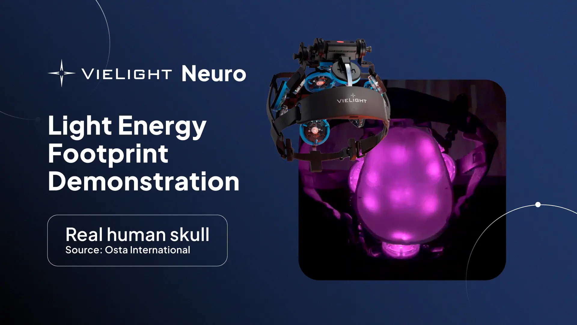
Seeing is Believing: CMOS Smartphone Photonic Detection
To make the diffusion concept visible, we ran simple visual experiments using a CMOS smartphone camera and a real human calvaria (see video below):
- Setup: The Vielight Neuro 4 and Vielight Neuro Pro 2 are positioned below a real human skull’s calvaria, which rests on top of it. A smartphone camera, sensitive enough to detect relative near‑infrared light despite typical IR filtering – captured trans‑bone light patterns.
- Observation: Each VieLED produces a vibrant, wide intensity field, not a narrow spot. Overlapping fields were evident as brighter, blended regions.
- Interpretation: The relative intensity maps match expectations from multiple scattering and interface redirection across bone, meningeal, and CSF boundaries.
What this is and isn’t: The smartphone method is a qualitative, relative visualization – useful for pattern‑tracking and comparative intensity across positions. It is not a calibrated dosimetry system and doesn’t replace formal optical modeling or in‑tissue fluence measurements.
A Quick Tour of the Physics (In Brief)
- Scalp & skull: High reduced scattering and modest absorption broaden and attenuate incident beams, creating diffuse halos.
- Dura/arachnoid/CSF: While CSF is comparatively low‑scattering, interfaces and surface irregularities (arachnoid, trabeculae, sulcal geometry) redirect and redistribute light, aiding lateral spread across adjacent gyri.
- Gray matter: Additional forward‑biased scattering continues to smooth and widen the footprint within cortex.
Together, these layers transform point‑like sources into distributed fields that can be stacked where we want emphasis (e.g., DMN hubs) while maintaining broad coverage elsewhere.
Limitations & Next Steps
- Qualitative visualization: CMOS camera capture provides relative intensity, influenced by sensor IR filtering and auto‑exposure. Future work can add spectral characterization and fixed‑exposure protocols.
- Heterogeneity: Skull thickness, diploë content, sinus cavities, and CSF thickness vary across individuals, subtly reshaping footprints. Ongoing Monte Carlo modeling and in‑vivo NIRS/NIRI can refine priors.
- Dosimetry bridge: Linking surface power, fluence rate at depth, and biologic response remains an active engineering task. Calibrated phantoms and paired imaging can tighten these relationships.
Conclusion
The Vielight Neuro VieLED architecture is deceptively simple: by leveraging tissue optics, it yields an effectively full‑transcranial energy footprint with a purposeful DMN bias. The intranasal channel completes the map by accessing ventral forebrain across the porous cribriform plate, creating a complementary dorsal‑to‑ventral pathway. The calvaria‑based visualizations make the physics tangible—five LEDs, one brain‑wide footprint, with DMN‑centered emphasis by design.
